- Home
- Arteriovenous Malformation Treatment
Arteriovenous Malformation Treatment in bhopal
Assessment and treatment of abnormal tangles of blood vessels in the brain.
Overview
An arteriovenous malformation (AVM) is an abnormal connection between arteries and veins that bypasses the usual network of tiny blood vessels called capillaries. A brain AVM can affect normal blood flow and may cause seizures, headaches, neurological symptoms, or bleeding. Some AVMs cause no noticeable symptoms and are discovered during investigations for another medical concern. Arteriovenous Malformation Treatment in Bhopal begins with understanding the AVM’s location, size, blood vessel structure, symptoms, and any history of bleeding. Treatment decisions also depend on the patient’s overall health and the potential risks of observation or intervention. Options may include monitoring, microsurgery, endovascular embolization, or stereotactic radiosurgery in selected cases. Not every AVM requires the same approach.
Dr. Harshad Patil is a neurosurgeon practising in Bhopal, Madhya Pradesh. He holds an MBBS from Terna Medical College & Hospital, Navi Mumbai, an MS in General Surgery from Dr. Vasantrao Pawar Medical College & Hospital, Nashik, and an M.Ch. in Neurosurgery from Sri Aurobindo Medical College and PG Institute, Indore. A neurosurgical consultation can help patients understand their scan reports, discuss possible management options, and prepare questions for their treating medical team.
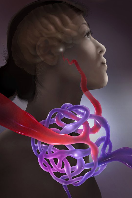
What Is an Arteriovenous Malformation?
Normally, arteries carry blood away from the heart, and veins return blood towards the heart. Between them, capillaries allow oxygen and nutrients to pass into surrounding tissues. An arteriovenous malformation (AVM) creates an abnormal connection between arteries and veins without the usual capillary network. This changes the way blood flows through the affected area. In a brain AVM, abnormal blood flow can place stress on blood vessels and may affect nearby brain tissue.
Some AVMs remain undetected for years. Others are discovered after a seizure, headache, neurological symptom, or bleeding event. Symptoms and risks vary considerably, so the diagnosis needs to be considered in the context of the individual patient. An AVM’s size alone does not determine the best management approach. Its position, blood vessel structure, drainage pattern, symptoms, and previous bleeding history can all influence treatment planning.
When considering Arteriovenous Malformation Treatment in Bhopal, it is important to understand that not every AVM requires surgery. In some cases, monitoring may be considered; in others, intervention may be recommended after careful assessment. An Arteriovenous Malformation Specialist in Bhopal can help patients understand their diagnosis and discuss the potential benefits, risks, and limitations of the available management options with their treating medical team.
Types and Locations of Arteriovenous Malformation Treatment in Bhopal
AVMs can occur in different parts of the body, but this page focuses on cerebral AVMs, which involve blood vessels in the brain.
Brain or cerebral AVMs
A brain AVM consists of an abnormal network of blood vessels within or around brain tissue. The precise anatomy varies from one patient to another. Some AVMs are near the brain’s surface, while others are located deeper within the brain.
Why location matters
An AVM’s location may influence the symptoms it causes and the risks associated with treatment. For example, an AVM close to areas responsible for speech, movement, vision, or coordination may require particularly careful planning. Deeper or more complex AVMs may present different treatment challenges from those closer to the brain’s surface. The treating team considers these details alongside the patient’s medical history and general health. The goal of assessment is to understand the potential risks of the AVM and compare them with the possible risks and benefits of each management option.
Symptoms and Emergency Warning Signs
Some brain AVMs cause no symptoms. Others may affect brain function or cause bleeding. Symptoms depend partly on the AVM’s location and whether a complication has occurred.
Possible symptoms include:
- Seizures: Unusual electrical activity in the brain may cause a seizure.
- Headaches: Some people experience recurring headaches, although headaches have many possible causes.
- Weakness or numbness: Symptoms may affect one part of the body, depending on the area involved.
- Speech difficulties: An AVM affecting relevant brain regions may be associated with trouble speaking or understanding language.
- Vision changes: Some patients may experience changes in vision.
- Balance or coordination problems: These may occur when areas involved in movement and balance are affected.
- Other neurological changes: Symptoms vary according to the AVM’s location and clinical effects.
These symptoms do not necessarily mean that a person has an AVM. Medical assessment is important to identify the cause.
Signs that require emergency care
Bleeding from a brain AVM can be serious and may cause sudden symptoms. Seek emergency care immediately if someone experiences:
- A sudden, severe headache.
- A seizure, especially a first seizure.
- New weakness or numbness.
- Difficulty speaking or understanding speech.
- Sudden loss of balance or major vision changes.
- Collapse or reduced consciousness.
Call 112 or seek immediate emergency medical care. Do not wait for a routine appointment, try to manage suspected brain bleeding at home, or delay emergency assessment to arrange an elective scan.
Causes and Risk Factors
Brain AVMs are generally believed to develop before birth or during early development. However, the precise cause of an individual AVM is not always known. Most brain AVMs are not explained by a simple lifestyle-related cause. Certain inherited conditions can be associated with abnormal blood vessel development, but this does not mean that every person with an AVM has an inherited disorder. An AVM may remain undetected for many years because it does not always produce symptoms. It may be discovered when imaging is performed for a seizure, headache, neurological change, or another medical concern. Patients should tell their clinician about relevant personal and family medical history, previous neurological symptoms, and any known inherited conditions. This information can help the treating team decide whether further evaluation is appropriate.
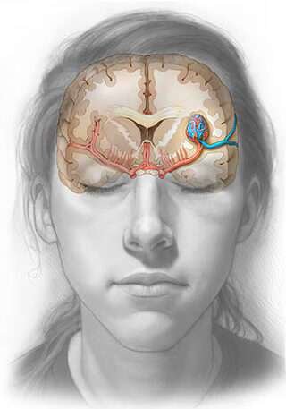
How Is a Brain AVM Diagnosed?
Diagnosis involves clinical assessment and appropriate imaging. The tests selected depend on the patient’s symptoms, medical history, and the information needed to understand the AVM.
MRI of the brain
Magnetic resonance imaging (MRI) produces detailed images of the brain. It can help identify an AVM and show its relationship to surrounding brain structures.
CT scan
A CT scan may be used when clinically indicated, particularly during emergency assessment if bleeding is suspected. It can provide important information about bleeding and other acute changes.
CT angiography and MR angiography
These tests help visualise blood vessels. They may provide information about the AVM’s structure, location, and blood supply.
Cerebral angiography
Cerebral angiography uses a catheter and contrast material to produce detailed images of blood vessels. It may be recommended when more precise information about the AVM’s anatomy is needed for treatment planning. Each investigation has a different purpose. The treating team determines which tests are appropriate and explains what the results mean. Sudden neurological symptoms require emergency assessment rather than waiting for elective imaging.
When considering Arteriovenous Malformation Treatment in Bhopal, patients should ask what their scan reports show, whether additional investigations are needed, and how the findings affect the possible management options.
Available AVM Treatment Options
Treatment is individualised. The decision depends on the AVM’s size, location, structure, symptoms, bleeding history, and the patient’s overall health. A medical team weighs the potential risks of leaving the AVM untreated against the risks of intervention.
Observation and monitoring
Selected AVMs may be monitored when the potential risks of intervention outweigh the expected benefits. Monitoring may involve clinical reviews and follow-up imaging according to the specialist team's recommendations. Observation does not mean ignoring the condition. Patients should understand their follow-up schedule, know which symptoms require urgent assessment, and attend recommended appointments.
Microsurgical removal
For selected AVMs, microsurgery may involve removing the abnormal blood vessel connection through an operation. Whether this is appropriate depends on the AVM's location, accessibility, complexity, and the potential effect of surgery on surrounding brain tissue. Surgical planning aims to balance the possibility of treating the AVM against the risk of neurological complications. Not every AVM is suitable for surgical removal.
Endovascular embolization
Endovascular embolization involves guiding a catheter through blood vessels and delivering a material into selected vessels to reduce or block blood flow through part of the AVM. Embolization may be used as part of a broader treatment plan or in selected cases. It does not necessarily eliminate the entire AVM, and the role of the procedure depends on its anatomy and the overall treatment strategy.
Stereotactic radiosurgery
Stereotactic radiosurgery delivers focused radiation to a selected AVM without an open surgical incision. The treatment works gradually, and the intended effect may take time to develop. The AVM does not necessarily disappear immediately after treatment. This approach is suitable only for selected cases. Patients should discuss the expected timeline, potential risks, and follow-up imaging requirements with the treating team.
Choosing an appropriate approach
AVM Treatment in Bhopal should be considered in the context of the patient's individual diagnosis and the options available through the responsible medical team. Monitoring, surgery, embolization, and radiosurgery are not interchangeable, and no single method is suitable for every AVM. A specialist assessment helps explain the reasoning behind a proposed approach and whether further evaluation by other specialists may be needed.
Available and Follow-Up
Risks and recovery vary according to the AVM’s location and complexity, the selected treatment, whether bleeding has occurred, and the patient’s general health. Possible risks associated with surgery or endovascular procedures may include bleeding, infection, blood vessel injury, stroke, or other neurological complications. These are potential complications, not inevitable outcomes. The treating team should explain the risks relevant to the patient’s case. After treatment, care may involve hospital monitoring, neurological assessment, medication management, follow-up imaging, and rehabilitation when needed. The recovery process differs between patients, so a fixed recovery time cannot be guaranteed. After stereotactic radiosurgery, the intended effect may take months or longer to develop, depending on the case. Follow-up imaging is important to assess the response and guide further care.
An Arteriovenous Malformation Treatment Specialist in Bhopal can help patients understand the follow-up recommendations and the questions they may wish to discuss with their treating team. Patients should take medicines as directed and should not stop or change prescribed medication without medical advice. Seek prompt medical attention if new or worsening neurological symptoms develop. Sudden severe headache, seizure, new weakness, difficulty speaking, collapse, or reduced consciousness should be treated as an emergency.
fAQ's
Frequently asked Questions❔
General information only. A consultation is needed for advice about an individual condition.
An arteriovenous malformation is an abnormal connection between arteries and veins that bypasses the usual capillary network. A brain AVM may cause seizures, headaches, neurological symptoms, or bleeding, although some AVMs cause no noticeable symptoms.
Arteriovenous Malformation Treatment in Bhopal may involve monitoring or an intervention such as surgery, embolization, or stereotactic radiosurgery in selected cases. The decision depends on the AVM's anatomy, symptoms, bleeding history, and the patient's overall health.
You can contact Dr. Harshad Patil on +91 75540 86000 to enquire about routine consultation arrangements. An Arteriovenous Malformation Doctor in Bhopal can discuss the available reports and possible next steps during a non-emergency consultation. Acute symptoms require emergency care instead.
Ask about the AVM's location and structure, the purpose of further tests, the potential risks of bleeding, and the benefits and limitations of monitoring or treatment. An Arteriovenous Malformation Specialist in Bhopal should help you understand the reasoning behind the proposed plan.
The assessment may include the AVM's location, size, blood vessel structure, symptoms, bleeding history, and the potential risks to nearby brain tissue. An Arteriovenous Malformation Surgeon in Bhopal can explain why surgery may or may not be appropriate and discuss alternatives.
AVM Treatment in Bhopal may involve monitoring, microsurgery, endovascular embolization, or stereotactic radiosurgery, depending on the individual case. These options have different benefits, limitations, and risks, so treatment should be discussed with the responsible medical team.
An Arteriovenous Malformation Treatment Specialist in Bhopal can help explain imaging results, treatment approaches, possible complications, and follow-up needs. Patients should ask whether further specialist opinions may be useful before deciding on a management plan.
No. Some AVMs may be monitored when intervention risks outweigh the expected benefits, while others may require treatment. The decision depends on the AVM's characteristics, symptoms, bleeding history, and the patient's overall health.